UW Neurological Surgery Recent PubMed Publications

Platelet Dynamics and Hemodynamics of Cerebral Aneurysms Treated with Flow-Diverting Stents.

5 years 6 months ago
Related Articles

Platelet Dynamics and Hemodynamics of Cerebral Aneurysms Treated with Flow-Diverting Stents.

Ann Biomed Eng. 2020 Jan;48(1):490-501

Authors: Marsh LMM, Barbour MC, Chivukula VK, Chassagne F, Kelly CM, Levy SH, Kim LJ, Levitt MR, Aliseda A

Abstract
Flow-diverting stents (FDS) are used to treat cerebral aneurysms. They promote the formation of a stable thrombus within the aneurysmal sac and, if successful, isolate the aneurysmal dome from mechanical stresses to prevent rupture. Platelet activation, a mechanism necessary for thrombus formation, is known to respond to biomechanical stimuli, particularly to the platelets' residence time and shear stress exposure. Currently, there is no reliable method for predicting FDS treatment outcomes, either a priori or after the procedure. Eulerian computational fluid dynamic (CFD) studies of aneurysmal flow have searched for predictors of endovascular treatment outcome; however, the hemodynamics of thrombus formation cannot be fully understood without considering the platelets' trajectories and their mechanics-triggered activation. Lagrangian analysis of the fluid mechanics in the aneurysmal vasculature provides novel metrics by tracking the platelets' residence time (RT) and shear history (SH). Eulerian and Lagrangian parameters are compared for 19 patient-specific cases, both pre- and post-treatment, to assess the degree of change caused by the FDS and subsequent treatment efficacy.

PMID: 31549329 [PubMed - indexed for MEDLINE]

Perinatal Breathing Patterns and Survival in Mice Born Prematurely and at Term.

5 years 6 months ago
Related Articles

Perinatal Breathing Patterns and Survival in Mice Born Prematurely and at Term.

Front Physiol. 2019;10:1113

Authors: Ramirez SC, Koschnitzky JE, Youngquist TM, Baertsch NA, Smith CV, Ramirez JM

Abstract
Infants born prematurely, often associated with maternal infection, frequently exhibit breathing instabilities that require resuscitation. We hypothesized that breathing patterns during the first hour of life would be predictive of survival in an animal model of prematurity. Using plethysmography, we measured breathing patterns during the first hour after birth in mice born at term (Term 19.5), delivered prematurely on gestational day 18.5 following administration of low-dose lipopolysaccharide (LPS; 0.14 mg/kg) to pregnant dams (LPS 18.5), or delivered on gestational day 18.7 or 17.5 by caesarian section (C-S 18.5 and C-S 17.5, respectively). Our experimental approach allowed us to dissociate effects caused by inflammation, from effects due to premature birth in the absence of an inflammatory response. C-S 17.5 mice did not survive, whereas mortality was not increased in C-S 18.5 mice. However, in premature pups born at the same gestational age (day 18.5) in response to maternal LPS injection, mortality was significantly increased. Overall, mice that survived had higher birth weights and showed eupneic or gasping activity that was able to transition to normal breathing. Some mice also exhibited a "saw tooth" breathing pattern that was able to transition into eupnea during the first hour of life. In contrast, mice that did not survive showed distinct, large amplitude, long-lasting breaths that occurred at low frequency and did not transition into eupnea. This breathing pattern was only observed during the first hour of life and was more prevalent in LPS 18.5 and C-S 18.5 mice. Indeed, breath tidal volumes were higher in inflammation-induced premature pups than in pups delivered via C-section at equivalent gestational ages, whereas breathing frequencies were low in both LPS-induced and C-section-induced premature pups. We conclude that a breathing pattern characterized by low frequency and large tidal volume is a predictor for the failure to survive, and that these characteristics are more often seen when prematurity occurs in the context of maternal inflammation. Further insights into the mechanisms that generate these breathing patterns and how they transition to normal breathing may facilitate development of novel strategies to manage premature birth in humans.

PMID: 31543825 [PubMed]

Estimated impact of supervised injection facilities on overdose fatalities and healthcare costs in New York City.

5 years 7 months ago
Related Articles

Estimated impact of supervised injection facilities on overdose fatalities and healthcare costs in New York City.

J Subst Abuse Treat. 2019 11;106:79-88

Authors: Behrends CN, Paone D, Nolan ML, Tuazon E, Murphy SM, Kapadia SN, Jeng PJ, Bayoumi AM, Kunins HV, Schackman BR

Abstract
BACKGROUND: The opioid epidemic in the United States has resulted in over 42,000 U.S. opioid overdose fatalities in 2016 alone. In New York City (NYC) opioid overdoses have reached a record high, increasing from 13.6 overdose deaths/100,000 to 19.9/100,000 from 2015 to 2016. Supervised injection facilities (SIFs) provide a hygienic, safe environment in which pre-obtained drugs can be consumed under clinical supervision to quickly reverse opioid overdoses. While SIFs have been implemented worldwide, none have been implemented to date in the United States. This study estimates the potential impact on opioid overdose fatalities and healthcare system costs of implementing SIFs in NYC.
METHODS: A deterministic model was used to project the number of fatal opioid overdoses avoided by implementing SIFs in NYC. Model inputs were from 2015 to 2016 NYC provisional overdose data (N = 1852) and the literature. Healthcare utilization and costs were estimated for fatal overdoses that would have been avoided from implementing one or more SIFs.
RESULTS: One optimally placed SIF is estimated to prevent 19-37 opioid overdose fatalities annually, representing a 6-12% decrease in opioid overdose mortality for that neighborhood; four optimally placed SIFs are estimated to prevent 68-131 opioid overdose fatalities. Opioid overdoses cost the NYC healthcare system an estimated $41 million per year for emergency medical services, emergency department visits, and hospitalizations. Implementing one SIF is estimated to save $0.8-$1.6 million, and four SIFs saves $2.9-$5.7 million in annual healthcare costs from opioid overdoses.
CONCLUSIONS: Implementing SIFs in NYC would save lives and healthcare system costs, although their overall impact may be limited depending on the geographic characteristic of the local opioid epidemic. In cities with geographically dispersed opioid epidemics such as NYC, multiple SIFs will be required to have a sizeable impact on the total number of opioid overdose fatalities occurring each year.

PMID: 31540615 [PubMed - indexed for MEDLINE]
Complete Microsurgical Resection of Large Retrochiasmatic Hypothalamic Craniopharyngioma by Transpetrosal Approach: 2-Dimensional Operative Video.

5 years 7 months ago
Related Articles

Complete Microsurgical Resection of Large Retrochiasmatic Hypothalamic Craniopharyngioma by Transpetrosal Approach: 2-Dimensional Operative Video.

Oper Neurosurg (Hagerstown). 2019 Sep 19;:

Authors: Zeeshan Q, Hernandez JPC, Yoh NT, Sekhar LN

Abstract
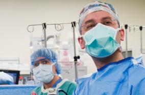
This two-dimensional video shows the technical nuances of complete microsurgical resection of a hypothalamic craniopharyngioma located in the retrochiasmatic region by the transpetrosal approach.  This 49-yr-old man presented with progressive fatigue, excessive sleepiness, and difficulty in vision in both eyes. He was found to have right CN 3 paralysis and bitemporal hemianopsia on neurological examination. Further workup revealed panhypopituitarism. Brain magnetic resonance imaging (MRI) demonstrated a large solid retrochiasmatic hypothalamic lesion with homogeneous contrast enhancement, measuring 2.1 × 2.6 × 2.4 cm. Optic chiasm was prefixed, and the tumor was just posterior to the pituitary stalk area. The preoperative differential diagnosis included hypothalamic astrocytoma, craniopharyngioma, germinoma, and histiocytosis. Because of the prefixed chiasm, a presigmoid, transpetrosal approach was performed. Our initial plan was a large biopsy, but based on frozen section histology, we decided to excise the tumor completely. The tumor had a pseudocapsule, which was firm and yellowish. It was debulked, dissected from the surrounding hypothalamus, and removed completely. The pituitary stalk was found at the anterior and inferior ends of the tumor and was preserved.  Postoperatively, the patient developed diabetes insipidus and requires desmopressin replacement, which was gradually tapered. For panhypopituitarism, he is receiving thyroxine, hydrocortisone, and testosterone.  Postoperatively, patient had an improvement in vision in his left eye and ptosis was improving in the right eye with mRs 1- at 10-wk follow-up.  An informed consent was obtained from the patient prior to the surgery, which included videotaping of the procedure and its distribution for educational purposes. All relevant patient identifiers have also been removed from the video and accompanying radiology slides.

PMID: 31538198 [PubMed - as supplied by publisher]

Neurosurgical management of Currarino syndrome: A case series and review of literature.

5 years 7 months ago
Related Articles

Neurosurgical management of Currarino syndrome: A case series and review of literature.

Surg Neurol Int. 2019;10:70

Authors: Tucker AM, Morgenstern P, Diaz D, Sedighim S, Shaul D, Sydorak R, Fedor M, Lee A, Hauptman J

Abstract
Background: The Currarino syndrome (CS), defined by the triad of anorectal malformations, sacral bone deformities, and presacral masses, is rare. There are few surgical series that discuss conservative management versus the surgical approaches to these lesions. Here, we describe utilizing a combined anterior and posterior approach for resecting these lesions in four patients.
Methods: Four patients with CS were treated with two-stage approaches performed by a multidisciplinary team, including pediatric neurosurgery and general surgery. The first anterior laparoscopic approach mobilized the presacral mass from its ventral attachments. The second posterior procedure detethered the spinal cord, repaired the dural defect, and facilitated removal of the presacral mass.
Results: Gross total resection of all four presacral masses was accomplished without intraoperative complication; all patients clinically improved.
Conclusion: The CS is characterized by a large presacral mass. Here, one must rule out malignancy and also consider diagnosis/resection due to the risks for malignant transformation. The operative approach we described in four patients utilized standard anterior mobilization of the mass, followed by posterior detethering, dural repair, and ultimate resection.

PMID: 31528408 [PubMed]

Mitotic Index Thresholds Do Not Predict Clinical Outcome for IDH-Mutant Astrocytoma.

5 years 7 months ago
Related Articles

Mitotic Index Thresholds Do Not Predict Clinical Outcome for IDH-Mutant Astrocytoma.

J Neuropathol Exp Neurol. 2019 11 01;78(11):1002-1010

Authors: Yoda RA, Marxen T, Longo L, Ene C, Wirsching HG, Keene CD, Holland EC, Cimino PJ

Abstract
Current histological grading recommendations for isocitrate dehydrogenase (IDH)-mutant astrocytoma are imprecise and not reliably predictive of patient outcome, while somatic copy number alterations are emerging as important prognostic biomarkers. One explanation for this relative underperformance of histological grading is that current criteria to distinguish World Health Organization (WHO) grade III anaplastic astrocytomas from lower-grade diffuse astrocytomas (WHO grade II) are vague ("increased mitotic activity"). This qualitative approach ensures diagnostic uncertainty and a broad "gray zone" where both diffuse and anaplastic designations can reasonably be assigned. Thus, we hypothesized that interobserver variability and lack of defined mitotic thresholds for IDH-mutant astrocytomas underlies poor predictive accuracy of current histologic grading approaches. To test this hypothesis, we quantified total mitotic figures and maximum mitotic activity per 10 high-powered fields in an institutional cohort of IDH-mutant astrocytomas. In our cohort, there was no mitotic activity threshold that was reflective of progression-free or overall survival (OS). Furthermore, in a multivariate Cox regression model consisting of mitotic activity, molecular markers, and clinical characteristics, only CDKN2A homozygous deletion was identified as a relevant variant for poor OS. We conclude that lack of defined mitotic figure thresholds may not contribute to underperformance of histological grading for IDH-mutant astrocytomas.

PMID: 31529048 [PubMed - indexed for MEDLINE]
Diabetes induces the activation of pro-ageing miR-34a in the heart, but has differential effects on cardiomyocytes and cardiac progenitor cells.

5 years 7 months ago
Related Articles

Diabetes induces the activation of pro-ageing miR-34a in the heart, but has differential effects on cardiomyocytes and cardiac progenitor cells.

Cell Death Differ. 2018 07;25(7):1336-1349

Authors: Fomison-Nurse I, Saw EEL, Gandhi S, Munasinghe PE, Van Hout I, Williams MJA, Galvin I, Bunton R, Davis P, Cameron V, Katare R

Abstract
Increased apoptosis and premature cellular ageing of the diabetic heart underpin the development of diabetic heart disease. The molecular mechanisms underlying these pathologies are still unclear. Here we determined the role of pro-senescence microRNA (miR)-34a in accelerating the ageing of the diabetic heart. RT-PCR analysis showed a significant increase in the level of circulating miR-34a from early stages in asymptomatic type-2 diabetic individuals compared to non-diabetic controls. We also observed significant upregulation of miR-34a in the type-2 human diabetic heart suggesting circulating miR-34a may be cardiac in origin. Moreover, western blot analysis identified marked downregulation of the pro-survival protein sirtuin 1 (SIRT1), a direct target of miR-34a. Analysis of cultured human adult cardiomyocytes exposed to high glucose and cardiac progenitor cells (CPCs) isolated from the diabetic heart confirmed significant upregulation of miR-34a and downregulation of SIRT1, associated with a marked increase in pro-apoptotic caspase-3/7 activity. Although therapeutic inhibition of miR-34a activity restored SIRT1 expression in both cardiomyocytes and CPCs, p53 expression was further upregulated in cardiomyocytes but conversely downregulated in CPCs. In spite of increased p53, miR-34a inhibition significantly reduced high glucose induced apoptotic cell death in cardiomyocytes. However, this effect was not observed in CPCs, which in fact showed reduced proliferation following miR-34a inhibition. Taken together, our results demonstrate upregulation of miR-34a in the diabetic heart and in the circulation from an early stage of the disease. However, inhibition of miR-34a activity has differential effects depending on the cell type, thereby warranting the need to eliminate off-target effects when introducing miR-based therapy.

PMID: 29302057 [PubMed - indexed for MEDLINE]
Surgery for very large and giant intracranial aneurysms: Results and complications.

5 years 7 months ago
Related Articles

Surgery for very large and giant intracranial aneurysms: Results and complications.

Neurol India. 2018 Nov-Dec;66(6):1741-1757

Authors: Zeeshan Q, Ghodke BV, Juric-Sekhar G, Barber JK, Kim LJ, Sekhar LN

Abstract
Background: Results of and the complications encountered during surgery for very large and giant intracranial aneurysms are illustrated.
Objective: To analyze a consecutive series of patients with very large and giant aneurysms treated with microsurgery.
Methods: This retrospective study included seventy six very large and giant aneurysms which were managed by clipping and bypass technique. Sixty two (82%) aneurysms were located in anterior circulation, and 14 (18%) aneurysms were located in posterior circulation. The bypasses performed included local bypasses, extra-intracranial bypasses, double bypasses and combination techniques of external carotid-internal carotid (EC-IC) bypass and local bypasses.
Results: 73 patients with 76 aneurysms were treated over 13 years. There were 44 very large and 32 giant aneurysms. Twenty-four patients presented with subarachnoid hemorrhage [SAH] (32%) while forty nine patients with 52 aneurysms (68%) were unruptured. These 73 patients underwent 63 bypass procedures with aneurysm occlusion and 13 clipping procedures. Out of 62 anterior circulation aneurysms, bypass surgery was performed in 49 patients while 13 underwent clipping. In posterior circulation aneurysms, all patients were treated with bypass procedures with proximal occlusion or trapping. In the ruptured group, 16 (67%) patients had postoperative modified Rankin Scale (mRs) 0-2, six patients (25%) had mRs 3-5, and two patients (8.4%) died. In the unruptured group, 45 patients (87%) had mRs 0-2, 3 patients (6%) had mRs 3-5, and four patients (7.6%) died.
Conclusions: In this large series of very large and giant aneurysms treated with microsurgical clipping and bypasses, excellent results were obtained in the long term, in regards to aneurysm occlusion, functional status, and graft patency. Our experience will be very useful to other neurosurgeons who treat these complex lesions.

PMID: 30504576 [PubMed - indexed for MEDLINE]

Timing of cranioplasty: a 10.75-year single-center analysis of 754 patients.

5 years 7 months ago
Related Articles

Timing of cranioplasty: a 10.75-year single-center analysis of 754 patients.

J Neurosurg. 2018 06;128(6):1648-1652

Authors: Morton RP, Abecassis IJ, Hanson JF, Barber JK, Chen M, Kelly CM, Nerva JD, Emerson SN, Ene CI, Levitt MR, Chowdhary MM, Ko AL, Chesnut RM

Abstract
OBJECTIVE Despite their technical simplicity, cranioplasty procedures carry high reported morbidity rates. The authors here present the largest study to date on complications after cranioplasty, focusing specifically on the relationship between complications and timing of the operation. METHODS The authors retrospectively reviewed all cranioplasty cases performed at Harborview Medical Center over the past 10.75 years. In addition to relevant clinical and demographic characteristics, patient morbidity and mortality data were abstracted from the electronic medical record. Cox proportional-hazards models were used to analyze variables potentially associated with the risk of infection, hydrocephalus, seizure, hematoma, and bone flap resorption. RESULTS Over the course of 10.75 years, 754 cranioplasties were performed at a single institution. Sixty percent of the patients who underwent these cranioplasties were male, and the median follow-up overall was 233 days. The 30-day mortality rate was 0.26% (2 cases, both due to postoperative epidural hematoma). Overall, 24.6% percent of the patients experienced at least 1 complication including infection necessitating explantation of the flap (6.6%), postoperative hydrocephalus requiring a shunt (9.0%), resorption of the flap requiring synthetic cranioplasty (6.3%), seizure (4.1%), postoperative hematoma requiring evacuation (2.3%), and other (1.6%). The rate of infection was significantly higher if the cranioplasty had been performed < 14 days after the initial craniectomy (p = 0.007, Holm-Bonferroni-adjusted p = 0.028). Hydrocephalus was significantly correlated with time to cranioplasty (OR 0.92 per 10-day increase, p < 0.001) and was most common in patients whose cranioplasty had been performed < 90 days after initial craniectomy. New-onset seizure, however, only occurred in patients who had undergone their cranioplasty > 90 days after initial craniectomy. Bone flap resorption was the least likely complication for patients whose cranioplasty had been performed between 15 and 30 days after initial craniectomy. Resorption was also correlated with patient age, with a hazard ratio of 0.67 per increase of 10 years of age (p = 0.001). CONCLUSIONS Cranioplasty performed between 15 and 30 days after initial craniectomy may minimize infection, seizure, and bone flap resorption, whereas waiting > 90 days may minimize hydrocephalus but may increase the risk of seizure.

PMID: 28799868 [PubMed - indexed for MEDLINE]
Cranial Chordoma: A New Preoperative Grading System.

5 years 7 months ago
Related Articles

Cranial Chordoma: A New Preoperative Grading System.

Neurosurgery. 2018 09 01;83(3):403-415

Authors: Brito da Silva H, Straus D, Barber JK, Rostomily RC, Ferreira M, Sekhar LN

Abstract
BACKGROUND: Chordomas are rare but challenging neoplasms involving the skull base. A preoperative grading system will be useful to identify both areas for treatment and risk factors, and correlate to the degree of resection, complications, and recurrence.
OBJECTIVE: To propose a new grading system for cranial chordomas designed by the senior author. Its purpose is to enable comparison of different tumors with a similar pathology to clivus chordoma, and statistically correlate with postoperative outcomes.
METHODS: The numerical grading system included tumor size, site of the tumor, vascular encasement, intradural extension, brainstem invasion, and recurrence of the tumor either after surgery or radiotherapy with a range of 2 to 25 points; it was used in 42 patients with cranial chordoma. The grading system was correlated with number of operations for resection, degree of resection, number and type of complications, recurrence, and survival.
RESULTS: We found 3 groups: low-risk 0 to 7 points, intermediate-risk 8 to 12 points, and high-risk ≥13 points in the grading system. The 3 groups were correlated with the following: extent of resection (partial, subtotal, or complete; P < .002); number of operative stages to achieve removal (P < .014); tumor recurrence (P = .03); postoperative Karnofsky Performance Status (P < .001); and with successful outcome (P = .005). The grading system itself correlated with the outcome (P = .005).
CONCLUSION: The proposed chordoma grading system can help surgeons to predict the difficulty of the case and know which areas of the skull base will need attention to plan further therapy.

PMID: 29126120 [PubMed - indexed for MEDLINE]

Post-Traumatic Hydrocephalus in Children: A Retrospective Study in 42 Pediatric Hospitals Using the Pediatric Health Information System.

5 years 7 months ago
Related Articles

Post-Traumatic Hydrocephalus in Children: A Retrospective Study in 42 Pediatric Hospitals Using the Pediatric Health Information System.

Neurosurgery. 2018 10 01;83(4):732-739

Authors: Bonow RH, Oron AP, Hanak BW, Browd SR, Chesnut RM, Ellenbogen RG, Vavilala MS, Rivara FP

Abstract
BACKGROUND: Post-traumatic hydrocephalus (PTH) is a potentially treatable cause of poor recovery from traumatic brain injury (TBI) that remains poorly understood, particularly among children.
OBJECTIVE: To better understand the risk factors for pediatric PTH using a large, multi-institutional database.
METHODS: We conducted a retrospective cohort study using administrative data from 42 pediatric hospitals participating in the Pediatric Health Information System. All patients ≤21 yr surviving a hospitalization with an International Classification of Diseases, Ninth Revision, Clinical Modification (ICD-9-CM) code for TBI were identified. The primary outcome was PTH, defined by an ICD-9-CM procedure code for surgical management of hydrocephalus within 6 mo. Data were analyzed using multivariable logistic regression.
RESULTS: We identified 91 583 patients ≤21 yr with TBI, 846 of whom developed PTH. Odds of PTH were significantly higher in children <1 yr compared to older age groups. A total of 48.7% of PTH cases were victims of abuse (adjusted odds ratio [aOR] 2.62, 95% confidence interval [CI] 2.16-3.18). PTH was more common after craniotomy (aOR 1.60, 95% CI 1.30-1.97). Craniectomy without early cranioplasty was associated with markedly increased odds of PTH (aOR 3.67, 95% CI 2.66-5.07), an effect not seen in those undergoing cranioplasty within 30 d (aOR 1.19, 95% CI 0.75-1.89).
CONCLUSION: PTH was seen in 0.9% of children who sustained a TBI and was more common in those <1 yr. Severe injury, abuse, and craniectomy with delayed cranioplasty were associated with greatly increased likelihood of PTH. Early cranioplasty in children who require craniectomy may reduce the risk for PTH.

PMID: 29029289 [PubMed - indexed for MEDLINE]

Dolichoectatic aneurysms of the vertebrobasilar system: clinical and radiographic factors that predict poor outcomes.

5 years 7 months ago
Related Articles

Dolichoectatic aneurysms of the vertebrobasilar system: clinical and radiographic factors that predict poor outcomes.

J Neurosurg. 2018 02;128(2):560-566

Authors: Xu DS, Levitt MR, Kalani MYS, Rangel-Castilla L, Mulholland CB, Abecassis IJ, Morton RP, Nerva JD, Siddiqui AH, Levy EI, Spetzler RF, Albuquerque FC, McDougall CG

Abstract
OBJECTIVE Fusiform dolichoectatic vertebrobasilar aneurysms are rare, challenging lesions. The natural history of these lesions and medium- and long-term patient outcomes are poorly understood. The authors sought to evaluate patient prognosis after diagnosis of fusiform dolichoectatic vertebrobasilar aneurysms and to identify clinical and radiographic predictors of neurological deterioration. METHODS The authors reviewed multiple, prospectively maintained, single-provider databases at 3 large-volume cerebrovascular centers to obtain data on patients with unruptured, fusiform, basilar artery dolichoectatic aneurysms diagnosed between January 1, 2000, and January 1, 2015. RESULTS A total of 50 patients (33 men, 17 women) were identified; mean clinical follow-up was 50.1 months and mean radiographic follow-up was 32.4 months. At last follow-up, 42% (n = 21) of aneurysms had progressed and 44% (n = 22) of patients had deterioration of their modified Rankin Scale scores. When patients were dichotomized into 2 groups- those who worsened and those who did not-univariate analysis showed 5 variables to be statistically significantly different: sex (p = 0.007), radiographic brainstem compression (p = 0.03), clinical posterior fossa compression (p < 0.001), aneurysmal growth on subsequent imaging (p = 0.001), and surgical therapy (p = 0.006). A binary logistic regression was then created to evaluate these variables. The only variable found to be a statistically significant predictor of clinical worsening was clinical symptoms of posterior fossa compression at presentation (p = 0.01). CONCLUSIONS Fusiform dolichoectatic vertebrobasilar aneurysms carry a poor prognosis, with approximately one-half of the patients deteriorating or experiencing progression of their aneurysm within 5 years. Despite being high risk, intervention-when carefully timed (before neurological decline)-may be beneficial in select patients.

PMID: 28387624 [PubMed - indexed for MEDLINE]

Reconstruction of 1,000 Projection Neurons Reveals New Cell Types and Organization of Long-Range Connectivity in the Mouse Brain.

5 years 7 months ago
Related Articles

Reconstruction of 1,000 Projection Neurons Reveals New Cell Types and Organization of Long-Range Connectivity in the Mouse Brain.

Cell. 2019 09 19;179(1):268-281.e13

Authors: Winnubst J, Bas E, Ferreira TA, Wu Z, Economo MN, Edson P, Arthur BJ, Bruns C, Rokicki K, Schauder D, Olbris DJ, Murphy SD, Ackerman DG, Arshadi C, Baldwin P, Blake R, Elsayed A, Hasan M, Ramirez D, Dos Santos B, Weldon M, Zafar A, Dudman JT, Gerfen CR, Hantman AW, Korff W, Sternson SM, Spruston N, Svoboda K, Chandrashekar J

Abstract
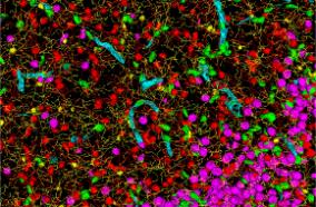
Neuronal cell types are the nodes of neural circuits that determine the flow of information within the brain. Neuronal morphology, especially the shape of the axonal arbor, provides an essential descriptor of cell type and reveals how individual neurons route their output across the brain. Despite the importance of morphology, few projection neurons in the mouse brain have been reconstructed in their entirety. Here we present a robust and efficient platform for imaging and reconstructing complete neuronal morphologies, including axonal arbors that span substantial portions of the brain. We used this platform to reconstruct more than 1,000 projection neurons in the motor cortex, thalamus, subiculum, and hypothalamus. Together, the reconstructed neurons constitute more than 85 meters of axonal length and are available in a searchable online database. Axonal shapes revealed previously unknown subtypes of projection neurons and suggest organizational principles of long-range connectivity.

PMID: 31495573 [PubMed - indexed for MEDLINE]

Acute kidney injury secondary to thrombotic microangiopathy associated with idiopathic hypereosinophilic syndrome: a case report and review of the literature.

5 years 7 months ago
Related Articles

Acute kidney injury secondary to thrombotic microangiopathy associated with idiopathic hypereosinophilic syndrome: a case report and review of the literature.

J Med Case Rep. 2019 Sep 05;13(1):281

Authors: Curras-Martin D, Patel S, Qaisar H, Mehandru SK, Masud A, Hossain MA, Lamba GS, Dounis H, Levitt M, Asif A

Abstract
BACKGROUND: Renal involvement in idiopathic hypereosinophilic syndrome is uncommon. The mechanism of kidney damage can be explained as occurring via two distinct pathways: (1) thromboembolic ischemic changes secondary to endocardial disruption mediated by eosinophilic cytotoxicity to the myocardium and (2) direct eosinophilic cytotoxic effect to the kidney.
CASE PRESENTATION: We present a case of a 63-year-old Caucasian man who presented to our hospital with 2 weeks of progressively generalized weakness. He was diagnosed with idiopathic hypereosinophilic syndrome with multiorgan involvement and acute kidney injury with biopsy-proven thrombotic microangiopathy. Full remission was achieved after 8 weeks of corticosteroid therapy.
CONCLUSION: Further studies are needed to investigate if age and absence of frank thrombocytopenia can serve as a prognostic feature of idiopathic hypereosinophilic syndrome, as seen in this case.

PMID: 31484586 [PubMed - indexed for MEDLINE]

Transradial intraoperative cerebral angiography: a multicenter case series and technical report.

5 years 7 months ago
Related Articles

Transradial intraoperative cerebral angiography: a multicenter case series and technical report.

J Neurointerv Surg. 2020 Feb;12(2):170-175

Authors: Osbun JW, Patel B, Levitt MR, Yahanda AT, Shah A, Dlouhy KM, Thatcher JP, Chicoine MR, Kim LJ, Zipfel GJ

Abstract
BACKGROUND: Use of the radial artery as an access site for neurointerventional procedures is gaining popularity after several studies in interventional cardiology have demonstrated superior patient safety, decreased length of stay, and patient preference compared with femoral artery access. The transradial approach has yet to be characterized for intraoperative cerebral angiography.
OBJECTIVE: To report a multicenter experience on the use of radial artery access in intraoperative cerebral angiography, including case series and discussion of technical nuances.
METHODS: 27 patients underwent attempted transradial cerebral angiography betweenMay 2017 and May 2019. Data were collected regarding technique, patient positioning, vessels selected, technical success rate, and access site complications.
RESULTS: 24 of the 27 patients (88.8%) underwent successful transradial intraoperative cerebral angiography. 18 patients (66.7%) were positioned supine, 6 patients (22.2%) were positioned prone, 1 patient (3.7%) was positioned lateral, and 2 patients (7.4%) were positioned three-quarters prone. A total of 31 vessels were selected including 13 right carotid arteries (8 common, 1 external, 4 internal), 11 left carotid arteries (9 common and 2 internal), and 6 vertebral arteries (5 right and 1 left). Two patients (7.4%) required conversion to femoral access in order to complete the intraoperative angiogram (1 due to arterial vasospasm and 1 due to inadvertent venous catheterization). One procedure (3.7%) was aborted because of inability to obtain the appropriate fluoroscopic views due to patient positioning. No patient experienced stroke, arterial dissection, or access site complication.
CONCLUSIONS: Transradial intraoperative cerebral angiography is safe and feasible with potential for improved operating room workflow ergonomics, faster patient mobility in the postoperative period, and reduced costs.

PMID: 31484699 [PubMed - indexed for MEDLINE]

Data for life cycle assessment of legume biorefining for alcohol.

5 years 7 months ago
Related Articles

Data for life cycle assessment of legume biorefining for alcohol.

Data Brief. 2019 Aug;25:104242

Authors: Lienhardt T, Black K, Saget S, Costa MP, Chadwick D, Rees R, Williams M, Spillane C, Iannetta P, Walker G, Styles D

Abstract
Benchmarking the environmental sustainability of alcohol produced from legume starch against alcohol produced from cereal grains requires considering of crop production, nutrient cycling and use of protein-rich co-products via life cycle assessment. This article describes the mass balance flows behind the life cycle inventories for gin produced from wheat and peas (Pisum sativum L.) in an associated article summarising the environmental footprints of wheat- and pea-gin [1], and also presents detailed supplementary results. Activity data were collected from interviews with actors along the entire gin value chain including a distillery manager and ingredient and packaging suppliers. Important fertiliser and animal-feed substitution effects of co-product use were derived using detailed information and models on nutrient flows and animal feed composition, along with linear optimisation modelling. Secondary data on environmental burdens of specific materials and processes were obtained from the Ecoinvent v3.4 life cycle assessment database. This article provides a basis for further quantitative evaluation of the environmental sustainability of legume-alcohol value chains.

PMID: 31485464 [PubMed]

Incidence and predictors of dural venous sinus pressure gradient in idiopathic intracranial hypertension and non-idiopathic intracranial hypertension headache patients: results from 164 cerebral venograms.

5 years 7 months ago
Related Articles

Incidence and predictors of dural venous sinus pressure gradient in idiopathic intracranial hypertension and non-idiopathic intracranial hypertension headache patients: results from 164 cerebral venograms.

J Neurosurg. 2017 02;126(2):347-353

Authors: Levitt MR, Hlubek RJ, Moon K, Kalani MY, Nakaji P, Smith KA, Little AS, Knievel K, Chan JW, McDougall CG, Albuquerque FC

Abstract
OBJECTIVE Cerebral venous pressure gradient (CVPG) from dural venous sinus stenosis is implicated in headache syndromes such as idiopathic intracranial hypertension (IIH). The incidence of CVPG in headache patients has not been reported. METHODS The authors reviewed all cerebral venograms with manometry performed for headache between January 2008 and May 2015. Patient demographics, headache etiology, intracranial pressure (ICP) measurements, and radiographic and manometric results were recorded. CVPG was defined as a difference ≥ 8 mm Hg by venographic manometry. RESULTS One hundred sixty-four venograms were performed in 155 patients. There were no procedural complications. Ninety-six procedures (58.5%) were for patients with IIH. The overall incidence of CVPG was 25.6% (42 of 164 procedures): 35.4% (34 of 96 procedures) in IIH patients and 11.8% (8 of 68 procedures) in non-IIH patients. Sixty procedures (36.6%) were performed in patients with preexisting shunts. Seventy-seven patients (49.7%) had procedures preceded by an ICP measurement within 4 weeks of venography, and in 66 (85.7%) of these patients, the ICP had been found to be elevated. CVPG was seen in 8.3% (n = 5) of the procedures in the 60 patients with a preexisting shunt and in 0% (n = 0) of the 11 procedures in the 77 patients with normal ICP (p < 0.001 for both). Noninvasive imaging (MR venography, CT venography) was assessed prior to venography in 112 (68.3%) of 164 cases, and dural venous sinus abnormalities were demonstrated in 73 (65.2%) of these cases; there was a trend toward CVPG (p = 0.07). Multivariate analysis demonstrated an increased likelihood of CVPG in patients with IIH (OR 4.97, 95% CI 1.71-14.47) and a decreased likelihood in patients with a preexisting shunt (OR 0.09, 95% CI 0.02-0.44). CONCLUSIONS CVPG is uncommon in IIH patients, rare in those with preexisting shunts, and absent in those with normal ICP.

PMID: 26967777 [PubMed - indexed for MEDLINE]

Meta-analysis and systematic review of risk factors for shunt dependency after aneurysmal subarachnoid hemorrhage.

5 years 7 months ago
Related Articles

Meta-analysis and systematic review of risk factors for shunt dependency after aneurysmal subarachnoid hemorrhage.

J Neurosurg. 2017 Feb;126(2):586-595

Authors: Wilson CD, Safavi-Abbasi S, Sun H, Kalani MY, Zhao YD, Levitt MR, Hanel RA, Sauvageau E, Mapstone TB, Albuquerque FC, McDougall CG, Nakaji P, Spetzler RF

Abstract
OBJECTIVE Aneurysmal subarachnoid hemorrhage (aSAH) may be complicated by hydrocephalus in 6.5%-67% of cases. Some patients with aSAH develop shunt dependency, which is often managed by ventriculoperitoneal shunt placement. The objectives of this study were to review published risk factors for shunt dependency in patients with aSAH, determine the level of evidence for each factor, and calculate the magnitude of each risk factor to better guide patient management. METHODS The authors searched PubMed and MEDLINE databases for Level A and Level B articles published through December 31, 2014, that describe factors affecting shunt dependency after aSAH and performed a systematic review and meta-analysis, stratifying the existing data according to level of evidence. RESULTS On the basis of the results of the meta-analysis, risk factors for shunt dependency included high Fisher grade (OR 7.74, 95% CI 4.47-13.41), acute hydrocephalus (OR 5.67, 95% CI 3.96-8.12), in-hospital complications (OR 4.91, 95% CI 2.79-8.64), presence of intraventricular blood (OR 3.93, 95% CI 2.80-5.52), high Hunt and Hess Scale score (OR 3.25, 95% CI 2.51-4.21), rehemorrhage (OR 2.21, 95% CI 1.24-3.95), posterior circulation location of the aneurysm (OR 1.85, 95% CI 1.35-2.53), and age ≥ 60 years (OR 1.81, 95% CI 1.50-2.19). The only risk factor included in the meta-analysis that did not reach statistical significance was female sex (OR 1.13, 95% CI 0.77-1.65). CONCLUSIONS The authors identified several risk factors for shunt dependency in aSAH patients that help predict which patients are likely to require a permanent shunt. Although some of these risk factors are not independent of each other, this information assists clinicians in identifying at-risk patients and managing their treatment.

PMID: 27035169 [PubMed - indexed for MEDLINE]

The Berlin International Consensus Meeting on Concussion in Sport.

5 years 7 months ago
Related Articles

The Berlin International Consensus Meeting on Concussion in Sport.

Neurosurgery. 2018 02 01;82(2):232-236

Authors: Davis GA, Ellenbogen RG, Bailes J, Cantu RC, Johnston KM, Manley GT, Nagahiro S, Sills A, Tator CH, McCrory P

Abstract
The Fifth International Conference on Concussion in Sport was held in Berlin in October 2016. A series of 12 questions and subquestions was developed and the expert panel members were required to perform a systematic review to answer each question. Following presentation at the Berlin meeting of the systematic review, poster abstracts and audience discussion, the summary Consensus Statement was produced. Further, a series of tools for the management of sport-related concussion was developed, including the Sport Concussion Assessment Tool Fifth edition (SCAT5), the Child SCAT5, and the Concussion Recognition Tool Fifth edition. This paper elaborates on this process, the outcomes, and explores the implications for neurosurgeons in the management of sport-related concussion.

PMID: 29106653 [PubMed - indexed for MEDLINE]

"university of washington"[affiliation] and neurological surge...: Latest results from PubMed
More posts about UW Neurological Surgery Recent PubMed Publications