UW Neurological Surgery Recent PubMed Publications

Author Correction: Genome-wide association and HLA fine-mapping studies identify risk loci and genetic pathways underlying allergic rhinitis.

5 years 8 months ago
Related Articles

Author Correction: Genome-wide association and HLA fine-mapping studies identify risk loci and genetic pathways underlying allergic rhinitis.

Nat Genet. 2018 Aug 16;:

Authors: Waage J, Standl M, Curtin JA, Jessen LE, Thorsen J, Tian C, Schoettler N, 23andMe Research Team, AAGC collaborators, Flores C, Abdellaoui A, Ahluwalia TS, Alves AC, Amaral AFS, Antó JM, Arnold A, Barreto-Luis A, Baurecht H, van Beijsterveldt CEM, Bleecker ER, Bonàs-Guarch S, Boomsma DI, Brix S, Bunyavanich S, Burchard EG, Chen Z, Curjuric I, Custovic A, den Dekker HT, Dharmage SC, Dmitrieva J, Duijts L, Ege MJ, Gauderman WJ, Georges M, Gieger C, Gilliland F, Granell R, Gui H, Hansen T, Heinrich J, Henderson J, Hernandez-Pacheco N, Holt P, Imboden M, Jaddoe VWV, Jarvelin MR, Jarvis DL, Jensen KK, Jónsdóttir I, Kabesch M, Kaprio J, Kumar A, Lee YA, Levin AM, Li X, Lorenzo-Diaz F, Melén E, Mercader JM, Meyers DA, Myers R, Nicolae DL, Nohr EA, Palviainen T, Paternoster L, Pennell CE, Pershagen G, Pino-Yanes M, Probst-Hensch NM, Rüschendorf F, Simpson A, Stefansson K, Sunyer J, Sveinbjornsson G, Thiering E, Thompson PJ, Torrent M, Torrents D, Tung JY, Wang CA, Weidinger S, Weiss S, Willemsen G, Williams LK, Ober C, Hinds DA, Ferreira MA, Bisgaard H, Strachan DP, Bønnelykke K

Abstract
In the version of this article initially published, in Fig. 3, the y-axis numbering did not match the log scale indicated in the axis label. The error has been corrected in the HTML and PDF version of the article.

PMID: 30116036 [PubMed - as supplied by publisher]

Defining the Rhythmogenic Elements of Mammalian Breathing.

5 years 8 months ago
Related Articles

Defining the Rhythmogenic Elements of Mammalian Breathing.

Physiology (Bethesda). 2018 09 01;33(5):302-316

Authors: Ramirez JM, Baertsch N

Abstract
Breathing's remarkable ability to adapt to changes in metabolic, environmental, and behavioral demands stems from a complex integration of its rhythm-generating network within the wider nervous system. Yet, this integration complicates identification of its specific rhythmogenic elements. Based on principles learned from smaller rhythmic networks of invertebrates, we define criteria that identify rhythmogenic elements of the mammalian breathing network and discuss how they interact to produce robust, dynamic breathing.

PMID: 30109823 [PubMed - indexed for MEDLINE]

Proteomic analysis of monolayer-integrated proteins on lipid droplets identifies amphipathic interfacial α-helical membrane anchors.

5 years 9 months ago
Related Articles

Proteomic analysis of monolayer-integrated proteins on lipid droplets identifies amphipathic interfacial α-helical membrane anchors.

Proc Natl Acad Sci U S A. 2018 08 28;115(35):E8172-E8180

Authors: Pataki CI, Rodrigues J, Zhang L, Qian J, Efron B, Hastie T, Elias JE, Levitt M, Kopito RR

Abstract
Despite not spanning phospholipid bilayers, monotopic integral proteins (MIPs) play critical roles in organizing biochemical reactions on membrane surfaces. Defining the structural basis by which these proteins are anchored to membranes has been hampered by the paucity of unambiguously identified MIPs and a lack of computational tools that accurately distinguish monolayer-integrating motifs from bilayer-spanning transmembrane domains (TMDs). We used quantitative proteomics and statistical modeling to identify 87 high-confidence candidate MIPs in lipid droplets, including 21 proteins with predicted TMDs that cannot be accommodated in these monolayer-enveloped organelles. Systematic cysteine-scanning mutagenesis showed the predicted TMD of one candidate MIP, DHRS3, to be a partially buried amphipathic α-helix in both lipid droplet monolayers and the cytoplasmic leaflet of endoplasmic reticulum membrane bilayers. Coarse-grained molecular dynamics simulations support these observations, suggesting that this helix is most stable at the solvent-membrane interface. The simulations also predicted similar interfacial amphipathic helices when applied to seven additional MIPs from our dataset. Our findings suggest that interfacial helices may be a common motif by which MIPs are integrated into membranes, and provide high-throughput methods to identify and study MIPs.

PMID: 30104359 [PubMed - indexed for MEDLINE]
Precardiac organoids form two heart fields via Bmp/Wnt signaling.

5 years 9 months ago
Related Articles

Precardiac organoids form two heart fields via Bmp/Wnt signaling.

Nat Commun. 2018 Aug 07;9(1):3140

Authors: Andersen P, Tampakakis E, Jimenez DV, Kannan S, Miyamoto M, Shin HK, Saberi A, Murphy S, Sulistio E, Chelko SP, Kwon C

Abstract
The discovery of the first heart field (FHF) and the second heart field (SHF) led us to understand how cardiac lineages and structures arise during development. However, it remains unknown how they are specified. Here, we generate precardiac spheroids with pluripotent stem cells (PSCs) harboring GFP/RFP reporters under the control of FHF/SHF markers, respectively. GFP+ cells and RFP+ cells appear from two distinct areas and develop in a complementary fashion. Transcriptome analysis shows a high degree of similarities with embryonic FHF/SHF cells. Bmp and Wnt are among the most differentially regulated pathways, and gain- and loss-of-function studies reveal that Bmp specifies GFP+ cells and RFP+ cells via the Bmp/Smad pathway and Wnt signaling, respectively. FHF/SHF cells can be isolated without reporters by the surface protein Cxcr4. This study provides novel insights into understanding the specification of two cardiac origins, which can be leveraged for PSC-based modeling of heart field/chamber-specific disease.

PMID: 30087351 [PubMed - in process]

Timely Hemodynamic Resuscitation and Outcomes in Severe Pediatric Traumatic Brain Injury: Preliminary Findings.

5 years 9 months ago
Related Articles

Timely Hemodynamic Resuscitation and Outcomes in Severe Pediatric Traumatic Brain Injury: Preliminary Findings.

Pediatr Emerg Care. 2018 May;34(5):325-329

Authors: Kannan N, Wang J, Mink RB, Wainwright MS, Groner JI, Bell MJ, Giza CC, Zatzick DF, Ellenbogen RG, Boyle LN, Mitchell PH, Rivara FP, Rowhani-Rahbar A, Vavilala MS, PEGASUS (Pediatric Guideline Adherence Outcomes) Study

Abstract
OBJECTIVES: Early resuscitation may improve outcomes in pediatric traumatic brain injury (TBI). We examined the association between timely treatment of hypotension and hypoxia during early care (prehospital or emergency department locations) and discharge outcomes in children with severe TBI.
METHODS: Hypotension was defined as systolic blood pressure less than 70 + 2 (age in years), and hypoxia was defined as PaO2 less than 60 mm Hg or oxygen saturation less than 90% in accordance with the 2003 Brain Trauma Foundation guidelines. Timely treatment of hypotension and hypoxia during early care was defined as the treatment within 30 minutes of a documented respective episode. Two hundred thirty-six medical records of children younger than 18 years with severe TBI from 5 regional pediatric trauma centers were examined. Main outcomes were in-hospital mortality and discharge Glasgow Outcome Scale (GOS) score.
RESULTS: Hypotension occurred in 26% (60/234) during early care and was associated with in-hospital mortality (23.3% vs 8.6%; P = 0.01). Timely treatment of hypotension during early care occurred in 92% (55/60) by use of intravenous fluids, blood products, or vasopressors and was associated with reduced in-hospital mortality [adjusted relative risk (aRR), 0.46; 95% confidence interval, 0.24-0.90] and less likelihood of poor discharge GOS (aRR, 0.54; 95% confidence interval, 0.39-0.76) when compared to children with hypotension who were not treated in a timely manner. Early hypoxia occurred in 17% (41/236) and all patients received timely oxygen treatment.
CONCLUSIONS: Timely resuscitation during early care was common and associated with lower in-hospital mortality and favorable discharge GOS in severe pediatric TBI.

PMID: 27387972 [PubMed - indexed for MEDLINE]
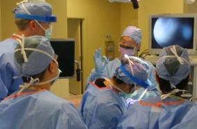
Study protocol: a cluster-randomized trial implementing Sustained Patient-centered Alcohol-related Care (SPARC trial).

5 years 9 months ago
Related Articles

Study protocol: a cluster-randomized trial implementing Sustained Patient-centered Alcohol-related Care (SPARC trial).

Implement Sci. 2018 08 06;13(1):108

Authors: Glass JE, Bobb JF, Lee AK, Richards JE, Lapham GT, Ludman E, Achtmeyer C, Caldeiro RM, Parrish R, Williams EC, Lozano P, Bradley KA

Abstract
BACKGROUND: Experts recommend that alcohol-related care be integrated into primary care (PC) to improve prevention and treatment of unhealthy alcohol use. However, few healthcare systems offer such integrated care. To address this gap, implementation researchers and clinical leaders at Kaiser Permanente Washington (KPWA) partnered to design a high-quality program of evidence-based care for unhealthy alcohol use: the Sustained Patient-centered Alcohol-related Care (SPARC) program. SPARC implements systems of clinical care designed to increase both prevention and treatment of unhealthy alcohol use. This clinical care for unhealthy alcohol use was implemented using three strategies: electronic health record (EHR) decision support, performance monitoring and feedback, and front-line support from external practice coaches with expertise in alcohol-related care ("SPARC implementation intervention" hereafter). The purpose of this report is to describe the protocol of the SPARC trial, a pragmatic, cluster-randomized, stepped-wedge implementation trial to evaluate whether the SPARC implementation intervention increased alcohol screening and brief alcohol counseling (so-called brief interventions), and diagnosis and treatment of alcohol use disorders (AUDs) in 22 KPWA PC clinics.
METHODS/DESIGN: The SPARC trial sample includes all adult patients who had a visit to any of the 22 primary care sites in the trial during the study period (January 1, 2015-July 31, 2018). The 22 sites were randomized to implement the SPARC program on different dates (in seven waves, approximately every 4 months). Primary outcomes are the proportion of patients with PC visits who (1) screen positive for unhealthy alcohol use and have documented brief interventions and (2) have a newly recognized AUD and subsequently initiate and engage in alcohol-related care. Main analyses compare the rates of these primary outcomes in the pre- and post-implementation periods, following recommended approaches for analyzing stepped-wedge trials. Qualitative analyses assess barriers and facilitators to implementation and required adaptations of implementation strategies.
DISCUSSION: The SPARC trial is the first study to our knowledge to use an experimental design to test whether practice coaches with expertise in alcohol-related care, along with EHR clinical decision support and performance monitoring and feedback to sites, increase both preventive care-alcohol screening and brief intervention-as well as diagnosis and treatment of AUDs.
TRIAL REGISTRATION: The trial is registered at ClinicalTrials.Gov: NCT02675777. Registered February 5, 2016, https://clinicaltrials.gov/ct2/show/NCT02675777 .

PMID: 30081930 [PubMed - indexed for MEDLINE]

Image-Guided, Asleep Deep Brain Stimulation.

5 years 9 months ago
Related Articles

Image-Guided, Asleep Deep Brain Stimulation.

Prog Neurol Surg. 2018;33:94-106

Authors: Ko AL, Burchiel KJ

Abstract
Deep brain stimulation (DBS) has become an established treatment for medically refractory movement disorders including Parkinson's disease, essential tremor, and dystonia. The field of DBS continues to evolve with advances in patient selection, target identification, electrode and pulse generator technology, and the development of more effective stimulation paradigms such as closed-loop stimulation. Furthermore, as the safety and efficacy of DBS improves through better hardware design and deeper understanding of its mechanisms of action, the indications for DBS will continue to expand to cover a wider range of disorders. Finally, the recent approval of MR-guided focused ultrasound for the treatment of essential tremor and potentially other movement disorders heralds a resurgence in lesion creation as a viable alternative to DBS for selected patients.

PMID: 29332076 [PubMed - indexed for MEDLINE]
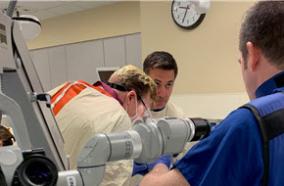
Predictors of infection after 754 cranioplasty operations and the value of intraoperative cultures for cryopreserved bone flaps.

5 years 9 months ago
Related Articles

Predictors of infection after 754 cranioplasty operations and the value of intraoperative cultures for cryopreserved bone flaps.

J Neurosurg. 2016 09;125(3):766-70

Authors: Morton RP, Abecassis IJ, Hanson JF, Barber J, Nerva JD, Emerson SN, Ene CI, Chowdhary MM, Levitt MR, Ko AL, Dellit TH, Chesnut RM

Abstract
OBJECTIVE The authors' aim was to report the largest study on predictors of infection after cranioplasty and to assess the predictive value of intraoperative bone flap cultures before cryopreservation. METHODS They retrospectively examined all cranioplasties performed between March 2004 and November 2014. Throughout this study period, the standard protocol during initial craniectomy was to obtain a culture swab of the extracted autologous bone flap (ABF)-prior to its placement in cytostorage-to screen for microbial contamination. Two consecutive protocols were employed for the use and interpretation of the intraoperative swab culture results: A) From March 2004 through June 2013, any culture-positive ABF (+ABF) was discarded and a custom synthetic prosthesis was implanted at the time of cranioplasty. B) From July 2013 through November 2014, any ABF with a skin flora organism was not discarded. Instead, cryopreservation was maintained and the +ABF was reimplanted after a 10-minute soak in bacitracin irrigation as well as a 3-minute soak in betadine. RESULTS Over the 10.75-year period, 754 cranioplasty procedures were performed. The median time from craniectomy to cranioplasty was 123 days. Median follow-up after cranioplasty was 237 days for protocol A and 225 days for protocol B. The overall infection rate after cranioplasty was 6.6% (50 cases) occurring at a median postoperative Day 31. Staphylococcus spp. were involved as the causative organisms in 60% of cases. Culture swabs taken at the time of initial craniectomy were available for 640 ABFs as 114 ABFs were not salvageable. One hundred twenty-six (20%) were culture positive. Eighty-nine +ABFs occurred during protocol A and were discarded in favor of a synthetic prosthesis at the time of cranioplasty, whereas 37 +ABFs occurred under protocol B and were reimplanted at the time of cranioplasty. Cranioplasty material did not affect the postcranioplasty infection rate. There was no significant difference in the infection rate among sterile ABFs (7%), +ABFs (8%), and synthetic prostheses (5.5%; p = 0.425). All 3 +ABF infections under protocol B were caused by organisms that differed from those in the original intraoperative bone culture from the initial craniectomy. A cranioplasty procedure ≤ 14 days after initial craniectomy was the only significant predictor of postcranioplasty infection (p = 0.007, HR 3.62). CONCLUSIONS Cranioplasty procedures should be performed at least 14 days after initial craniectomy to minimize infection risk. Obtaining intraoperative bone cultures at the time of craniectomy in the absence of clinical infection should be discontinued as the culture results were not a useful predictor of postcranioplasty infection and led to the unnecessary use of synthetic prostheses and increased health care costs.

PMID: 26771856 [PubMed - indexed for MEDLINE]

Subtraction multiphase CT angiography: A new technique for faster detection of intracranial arterial occlusions.

5 years 9 months ago
Related Articles

Subtraction multiphase CT angiography: A new technique for faster detection of intracranial arterial occlusions.

Eur Radiol. 2018 Apr;28(4):1731-1738

Authors: Byrne D, Walsh JP, Sugrue G, Stanley E, Marnane M, Walsh CD, Kelly P, Murphy S, Kavanagh EC, MacMahon PJ

Abstract
OBJECTIVE: To describe and evaluate a novel technical development to improve detection of intracranial vessel occlusions using multiphase CT angiography (MPCTA).
MATERIALS AND METHODS: The institutional ethics committee approved the study. Fifty patients (30 consecutive distal (M2 or smaller) anterior circulation occlusions, ten M1 occlusions, ten cases without occlusion) presenting with suspected AIS who underwent MPCTA were included. Post-processing of MPCTA studies created "subtraction" and "delayed enhancement" (DE) datasets. Initially, non-contrast CT and MPCTA studies for each patient were evaluated. Readers' confidence, speed and sensitivity of detection of intracranial vessel occlusions were recorded. After an interval of at least 4 weeks, readers were provided with post-processed images and studies were re-evaluated.
RESULTS: While the sensitivity of detection of intracranial vessel occlusions was equal for both conventional MPCTA and subMPCTA, the mean time taken to identify a vessel occlusion decreased by 64 % using subMPCTA (16 s vs. 45 s with conventional MPCTA) (p<0.001). In addition, confidence in interpretation improved (from 4.4 to 4.9) using subMPCTA (p<0.001).
CONCLUSION: SubMPCTA is a novel technique that aids in identifying small intracranial vessel occlusions in the suspected AIS patient. SubMPCTA increases confidence in interpretation and reduces the time taken to detect intracranial vessel occlusions.
KEY POINTS: • SubMPCTA processes MPCTA data to better demonstrate intracranial arterial occlusions. • SubMPCTA increases confidence and speed of interpretation of MPCTA studies. • SubMPCTA may aid in rapidly differentiating acute ischaemic stroke from stroke mimics.

PMID: 29134350 [PubMed - indexed for MEDLINE]

Anesthetics Have Different Effects on the Electrocorticographic Spectra of Wild-type and Mitochondrial Mutant Mice.

5 years 9 months ago
Related Articles

Anesthetics Have Different Effects on the Electrocorticographic Spectra of Wild-type and Mitochondrial Mutant Mice.

Anesthesiology. 2018 10;129(4):744-755

Authors: Carspecken CW, Chanprasert S, Kalume F, Sedensky MM, Morgan PG

Abstract
WHAT WE ALREADY KNOW ABOUT THIS TOPIC: WHAT THIS ARTICLE TELLS US THAT IS NEW: BACKGROUND:: Knockout of the mitochondrial protein Ndufs4 (Ndufs4[KO]) in mice causes hypersensitivity to volatile anesthetics but resistance to ketamine. The authors hypothesized that electrocorticographic changes underlying the responses of Ndufs4(KO) to volatile anesthetics and to ketamine would be similar in mutant and control mice.
METHODS: Electrocorticographic recordings at equipotent volatile anesthetic concentrations were compared between genotypes. In separate studies, control and cell type-specific Ndufs4(KO) mice were anesthetized with intraperitoneal ketamine to determine their ED50s.
RESULTS: Ndufs4 (KO) did not differ from controls in baseline electrocorticography (N = 5). Compared to baseline, controls exposed to isoflurane (EC50) lost power (expressed as mean baseline [µV/Hz]; mean isoflurane [µV/Hz]) in delta (2.45; 0.50), theta (1.41; 0.16), alpha (0.23; 0.05), beta (0.066; 0.016), and gamma (0.020; 0.005) frequency bands (N = 5). Compared to baseline, at their isoflurane EC50, Ndufs4(KO) maintained power in delta (1.08; 1.38), theta (0.36; 0.26), and alpha (0.09; 0.069) frequency bands but decreased in beta (0.041; 0.023) and gamma (0.020; 0.0068) frequency bands (N = 5). Similar results were seen for both genotypes in halothane. Vesicular glutamate transporter 2 (VGLUT2)-specific Ndufs4(KO) mice were markedly resistant to ketamine (ED50; 125 mg/kg) compared to control mice (ED50; 75 mg/kg; N = 6). At their respective ED95s for ketamine, mutant (N = 5) electrocorticography spectra showed a decrease in power in the beta (0.040; 0.020) and gamma (0.035; 0.015) frequency bands not seen in controls (N = 7).
CONCLUSIONS: Significant differences exist between the electrocorticographies of mutant and control mice at equipotent doses for volatile anesthetics and ketamine. The energetic state specifically of excitatory neurons determines the behavioral response to ketamine.

PMID: 30074932 [PubMed - indexed for MEDLINE]

Risk of Major Bleeding with Ibrutinib.

5 years 9 months ago
Related Articles

Risk of Major Bleeding with Ibrutinib.

Clin Lymphoma Myeloma Leuk. 2018 11;18(11):755-761

Authors: Mock J, Kunk PR, Palkimas S, Sen JM, Devitt M, Horton B, Portell CA, Williams ME, Maitland H

Abstract
BACKGROUND: The Bruton tyrosine kinase inhibitor, ibrutinib, is an effective therapy against mature B-cell malignancies. Although generally well tolerated, serious bleeding emerged during developmental clinical trials as an unexpected, although uncommon, adverse event. As the use of ibrutinib increases outside of the clinical trial setting and in patients with more comorbidities, the rate of major bleeding could be greater.
MATERIALS AND METHODS: A retrospective analysis the data from all patients at our center and its regional clinics who had been prescribed ibrutinib from January 2012 to May 2016 were reviewed for demographic data, comorbid illnesses, bleeding events, and concurrent medications.
RESULTS: We identified 70 patients. Bleeding of any grade occurred in 56% of patients, mostly grade 1 to 2 bruising and epistaxis. Major bleeding, defined as grade ≥ 3, occurred in 19% of patients, greater than previously reported. Anemia (hemoglobin < 12 g/dL; hazard ratio [HR], 5.0; 95% confidence interval [CI], 1.4-18.2; P = .02) and an elevated international normalized ratio (> 1.5; HR, 9.5; 95% CI, 2.7-33.5; P < .01) at ibrutinib initiation were associated with an increased risk of major bleeding. Of those with major bleeding, most patients were also taking an antiplatelet agent (70%), an anticoagulant (17%), or a CYP 3A4 inhibitor (7%), with 13% taking both antiplatelet and anticoagulant medications. The use of both antiplatelet and anticoagulant therapy significantly increased the risk of a major bleed event (HR, 19.2; 95% CI, 2.3-166.7; P < .01).
CONCLUSION: The results of the present study have demonstrated a greater rate of major bleeding with ibrutinib use in a standard clinical setting than previously reported. Patients with anemia or an elevated international normalized ratio or requiring anticoagulant and/or antiplatelet medications during ibrutinib therapy have a significantly increased risk of major bleeding. Careful consideration of the risks and benefits for this population is needed. The combination of antiplatelet and anticoagulation medications with ibrutinib therapy is of particular concern.

PMID: 30077698 [PubMed - indexed for MEDLINE]

Approaches to closed-loop deep brain stimulation for movement disorders.

5 years 9 months ago
Related Articles

Approaches to closed-loop deep brain stimulation for movement disorders.

Neurosurg Focus. 2018 08;45(2):E2

Authors: Kuo CH, White-Dzuro GA, Ko AL

Abstract
OBJECTIVE Deep brain stimulation (DBS) is a safe and effective therapy for movement disorders, such as Parkinson's disease (PD), essential tremor (ET), and dystonia. There is considerable interest in developing "closed-loop" DBS devices capable of modulating stimulation in response to sensor feedback. In this paper, the authors review related literature and present selected approaches to signal sources and approaches to feedback being considered for deployment in closed-loop systems. METHODS A literature search using the keywords "closed-loop DBS" and "adaptive DBS" was performed in the PubMed database. The search was conducted for all articles published up until March 2018. An in-depth review was not performed for publications not written in the English language, nonhuman studies, or topics other than Parkinson's disease or essential tremor, specifically epilepsy and psychiatric conditions. RESULTS The search returned 256 articles. A total of 71 articles were primary studies in humans, of which 50 focused on treatment of movement disorders. These articles were reviewed with the aim of providing an overview of the features of closed-loop systems, with particular attention paid to signal sources and biomarkers, general approaches to feedback control, and clinical data when available. CONCLUSIONS Closed-loop DBS seeks to employ biomarkers, derived from sensors such as electromyography, electrocorticography, and local field potentials, to provide real-time, patient-responsive therapy for movement disorders. Most studies appear to focus on the treatment of Parkinson's disease. Several approaches hold promise, but additional studies are required to determine which approaches are feasible, efficacious, and efficient.

PMID: 30064321 [PubMed - indexed for MEDLINE]

Anticipating the Direction of Soccer Penalty Shots Depends on the Speed and Technique of the Kick.

5 years 9 months ago
Related Articles

Anticipating the Direction of Soccer Penalty Shots Depends on the Speed and Technique of the Kick.

Sports (Basel). 2018 Jul 29;6(3):

Authors: Hunter AH, Murphy SC, Angilletta MJ, Wilson RS

Abstract
To succeed at a sport, athletes must manage the biomechanical trade-offs that constrain their performance. Here, we investigate a previously unknown trade-off in soccer: how the speed of a kick makes the outcome more predictable to an opponent. For this analysis, we focused on penalty kicks to build on previous models of factors that influence scoring. More than 700 participants completed an online survey, watching videos of penalty shots from the perspective of a goalkeeper. Participants (ranging in soccer playing experience from never played to professional) watched 60 penalty kicks, each of which was occluded at a particular moment (-0.4 s to 0.0 s) before the kicker contacted the ball. For each kick, participants had to predict shot direction toward the goal (left or right). As expected, predictions became more accurate as time of occlusion approached ball contact. However, the effect of occlusion was more pronounced when players kicked with the side of the foot than when they kicked with the top of the foot (instep). For side-foot kicks, the direction of shots was predicted more accurately for faster kicks, especially when a large portion of the kicker's approach was presented. Given the trade-off between kicking speed and directional predictability, a penalty kicker might benefit from kicking below their maximal speed.

PMID: 30060599 [PubMed]

Early Response Assessment in Pancreatic Ductal Adenocarcinoma Through Integrated PET/MRI.

5 years 9 months ago
Related Articles

Early Response Assessment in Pancreatic Ductal Adenocarcinoma Through Integrated PET/MRI.

AJR Am J Roentgenol. 2018 11;211(5):1010-1019

Authors: Wang ZJ, Behr S, Consunji MV, Yeh BM, Ohliger MA, Gao K, Ko AH, Cinar P, Tempero MA, Collisson EA

Abstract
OBJECTIVE: The purpose of this study is to investigate early changes in 18F-FDG PET/MRI metrics after treatment in patients with advanced pancreatic ductal adenocarcinoma (PDAC) and to correlate those changes with eventual tumor response at standard-of-care CT.
SUBJECTS AND METHODS: Thirteen patients with advanced PDAC underwent integrated FDG PET/MRI before and 4 weeks after treatment initiation. Patients were classified as responders or nonresponders according to Response Evaluation Criteria in Solid Tumors (RECIST) 1.1 at subsequent CT performed 8-12 weeks after treatment initiation. Changes in the primary tumor's maximum standardized uptake value (SUVmax), metabolic tumor volume (MTV), and total lesion glycolysis (TLG) determined at PET and apparent diffusion coefficient (ADC) determined at DWI at 4 weeks were compared between responders and nonresponders.
RESULTS: Seven patients had a partial response according to RECIST, and six did not. Responders displayed significantly greater decreases in MTV (p = 0.003) and TLG (p = 0.006) in the primary pancreatic tumor at 4 weeks. Responders also displayed a greater increase in the mean (p = 0.004) and minimum (p = 0.024) ADC of the primary tumors. Tumor size change at 4 weeks was not significantly different between responders and nonresponders (p = 0.11). PET responders enjoyed longer progression-free survival (PFS) (p = 0.0004) and overall survival (OS) (p = 0.013) than did nonresponders, using either a 60% reduction in MTV or 65% reduction in TLG as a threshold. MRI responders had significantly longer PFS (p = 0.0002) and OS (p = 0.027) than did nonresponders, using a 20% increase in either mean or minimum ADC as a threshold.
CONCLUSION: Integrated PET/MRI can provide early response assessment in patients with advanced PDAC, thus potentially allowing early treatment adaptation in nonresponders.

PMID: 30063366 [PubMed - indexed for MEDLINE]

Plasmodium 18S rRNA of intravenously administered sporozoites does not persist in peripheral blood.

5 years 9 months ago
Related Articles

Plasmodium 18S rRNA of intravenously administered sporozoites does not persist in peripheral blood.

Malar J. 2018 Jul 27;17(1):275

Authors: Murphy SC, Ishizuka AS, Billman ZP, Olsen TM, Seilie AM, Chang M, Smith N, Chuenchob V, Chakravarty S, Sim BKL, Kappe SHI, Hoffman SL, Seder RA

Abstract
BACKGROUND: Plasmodium 18S rRNA is a biomarker used to monitor blood-stage infections in malaria clinical trials. Plasmodium sporozoites also express this biomarker, and there is conflicting evidence about how long sporozoite-derived 18S rRNA persists in peripheral blood. If present in blood for an extended timeframe, sporozoite-derived 18S rRNA could complicate use as a blood-stage biomarker.
METHODS: Blood samples from Plasmodium yoelii infected mice were tested for Plasmodium 18S rRNA and their coding genes (rDNA) using sensitive quantitative reverse transcription PCR and quantitative PCR assays, respectively. Blood and tissues from Plasmodium falciparum sporozoite (PfSPZ)-infected rhesus macaques were similarly tested.
RESULTS: In mice, when P. yoelii sporozoite inoculation and blood collection were performed at the same site (tail vein), low level rDNA positivity persisted for 2 days post-infection. Compared to intact parasites with high rRNA-to-rDNA ratios, this low level positivity was accompanied by no increase in rRNA-to-rDNA, indicating detection of residual, non-viable parasite rDNA. When P. yoelii sporozoites were administered via the retro-orbital vein and blood sampled by cardiac puncture, neither P. yoelii 18S rRNA nor rDNA were detected 24 h post-infection. Similarly, there was no P. falciparum 18S rRNA detected in blood of rhesus macaques 3 days after intravenous injection with extremely high doses of PfSPZ. Plasmodium 18S rRNA in the rhesus livers increased by approximately 101-fold from 3 to 6 days post infection, indicating liver-stage proliferation.
CONCLUSIONS: Beyond the first few hours after injection, sporozoite-derived Plasmodium 18S rRNA was not detected in peripheral blood. Diagnostics based on 18S rRNA are unlikely to be confounded by sporozoite inocula in human clinical trials.

PMID: 30053881 [PubMed - in process]

Role of a conserved glutamine in the function of voltage-gated Ca2+ channels revealed by a mutation in human CACNA1D.

5 years 9 months ago
Related Articles

Role of a conserved glutamine in the function of voltage-gated Ca2+ channels revealed by a mutation in human CACNA1D.

J Biol Chem. 2018 09 14;293(37):14444-14454

Authors: Garza-Lopez E, Lopez JA, Hagen J, Sheffer R, Meiner V, Lee A

Abstract
Voltage-gated Cav Ca2+ channels play crucial roles in regulating gene transcription, neuronal excitability, and synaptic transmission. Natural or pathological variations in Cav channels have yielded rich insights into the molecular determinants controlling channel function. Here, we report the consequences of a natural, putatively disease-associated mutation in the CACNA1D gene encoding the pore-forming Cav1.3 α1 subunit. The mutation causes a substitution of a glutamine residue that is highly conserved in the extracellular S1-S2 loop of domain II in all Cav channels with a histidine and was identified by whole-exome sequencing of an individual with moderate hearing impairment, developmental delay, and epilepsy. When introduced into the rat Cav1.3 cDNA, Q558H significantly decreased the density of Ca2+ currents in transfected HEK293T cells. Gating current analyses and cell-surface biotinylation experiments suggested that the smaller current amplitudes caused by Q558H were because of decreased numbers of functional Cav1.3 channels at the cell surface. The substitution also produced more sustained Ca2+ currents by weakening voltage-dependent inactivation. When inserted into the corresponding locus of Cav2.1, the substitution had similar effects as in Cav1.3. However, the substitution introduced in Cav3.1 reduced current density, but had no effects on voltage-dependent inactivation. Our results reveal a critical extracellular determinant of current density for all Cav family members and of voltage-dependent inactivation of Cav1.3 and Cav2.1 channels.

PMID: 30054272 [PubMed - indexed for MEDLINE]

Vitamin D Levels in Patients with Colorectal Cancer Before and After Treatment Initiation.

5 years 9 months ago
Related Articles

Vitamin D Levels in Patients with Colorectal Cancer Before and After Treatment Initiation.

J Gastrointest Cancer. 2019 Dec;50(4):769-779

Authors: Savoie MB, Paciorek A, Zhang L, Van Blarigan EL, Sommovilla N, Abrams D, Atreya CE, Bergsland EK, Chern H, Kelley RK, Ko A, Laffan A, Sarin A, Varma MG, Venook AP, Van Loon K

Abstract
PURPOSE: We aimed to described 25-hydroxyvitamin D [25(OH)D] levels in newly diagnosed colorectal cancer (CRC) patients and to re-evaluate levels after chemotherapy.
METHODS: Permanent residents of the San Francisco Bay Area with a new CRC diagnosis of any stage were recruited prior to any non-surgical therapy. Serum 25(OH)D levels were measured at time of diagnosis and 6-month follow-up. Supplement use was not restricted. The primary endpoint was the frequency of vitamin D deficiency in patients with newly diagnosed CRC of all stages. The Kruskal-Wallis and Spearman correlation tests were used to evaluate associations of patient characteristics with 25(OH)D levels.
RESULTS: Median 25(OH)D level at baseline was 27.0 ng/mL (range 7.2, 59.0); 65% of patients had insufficient levels (25(OH)D < 30 ng/mL) (n = 94). Race, disease stage, multivitamin use, vitamin D supplementation, and county of residence were associated with baseline 25(OH)D levels (P < 0.05). The median change in 25(OH)D from baseline to 6 months was - 0.7 ng/mL [- 19.4, 51.7] for patients treated with chemotherapy (n = 58) and 1.6 ng/mL [- 6.4, 33.2] for patients who did not receive chemotherapy (n = 19) (P = 0.26). For patients who received vitamin D supplementation during chemotherapy, the median 25(OH)D change was 8.3 ng/mL [- 7.6, 51.7] versus - 1.6 [- 19.4, 24.3] for chemotherapy patients who did not take vitamin D supplements (P = 0.02).
CONCLUSION: Among patients with a new diagnosis of CRC, most patients were found to have 25(OH)D levels consistent with either deficiency or insufficiency. In the subset of patients who received chemotherapy and took a vitamin D supplement, serum 25(OH)D levels increased, suggesting that vitamin D repletion is a feasible intervention during chemotherapy.

PMID: 30058032 [PubMed - indexed for MEDLINE]

Intra-abdominal complications following intestinal anastomoses by suture and staple techniques in dogs.

5 years 9 months ago
Related Articles

Intra-abdominal complications following intestinal anastomoses by suture and staple techniques in dogs.

J Am Vet Med Assoc. 2018 Aug 15;253(4):437-443

Authors: DePompeo CM, Bond L, George YE, Mezzles MJ, Brourman JD, Chandler JC, Murphy SM, Pike F, Mason DR

Abstract
OBJECTIVE To compare the incidence of intra-abdominal complications in dogs following resection and functional end-to-end stapled anastomosis (FEESA) versus anastomosis with an end-to-end sutured technique for treatment of enteric lesions. DESIGN Multicenter, retrospective descriptive cohort study. ANIMALS 180 dogs. PROCEDURES Medical records of dogs undergoing intestinal resection and anastomosis at 3 nonaffiliated private practice specialty centers were retrospectively reviewed. Preoperative clinical variables, indication for surgery, surgical technique (sutured end-to-end anastomosis vs FEESA), and evidence of postoperative anastomosis site leakage (dehiscence) were recorded. Variables of interest were analyzed for associations with dehiscence. RESULTS Dehiscence rates of sutured and stapled anastomoses were 12 of 93 (13%) and 4 of 87 (5%), respectively; odds of postoperative dehiscence were significantly lower for dogs with FEESAs than for dogs with sutured anastomoses (OR, 0.28; 95% confidence interval, 0.09 to 0.94). Among dogs that underwent surgery for treatment of intestinal dehiscence after surgery at another facility, subsequent dehiscence developed in 3 of 5 with sutured anastomoses and 0 of 11 with stapled anastomoses. Dehiscence rates varied significantly among clinics. No other variable was associated with risk of dehiscence. Eleven of 16 dogs with dehiscence were euthanized without additional surgery. Impaction at the anastomosis site was identified months or years after surgery in 3 dogs (4 anastomosis sites) that had FEESAs. CONCLUSIONS AND CLINICAL RELEVANCE Odds for dehiscence were significantly greater for sutured end-to-end anastomoses than FEESAs, and dogs undergoing surgery for previous dehiscence were significantly more likely to experience a subsequent dehiscence with a sutured anastomosis. However, variability of procedure types and dehiscence rates among clinics suggested further research is needed to confirm these findings. Obstruction at the anastomosis site was identified as a potential long-term complication of FEESA.

PMID: 30058972 [PubMed - indexed for MEDLINE]

"university of washington"[affiliation] and neurological surge...: Latest results from PubMed
More posts about UW Neurological Surgery Recent PubMed Publications