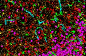
Normal aging can result in 20–30% reduction of microvasculature resulting in the cerebral cortex. Significant drop in capillary density as well as angiogenesis capabilities have been detected in both grey and white matter within the brain. This phenomenon of reduction in blood vessels density and blood supply in normal aging is thought to underlie the “sensitive” nature of aging brain to ischemic injuries. Interestingly, according to estimates from the American Academy of Orthopedic Surgery, spinal stenosis, narrowing of the spinal cord, affects 8 to 11 percent of the population in the United States, with nearly 2.4 million Americans are expected to be affected by 2021. Indeed, age is a significant risk factor for spinal stenosis, underscored by one study evaluating the spinal cord of ~ 450 asymptomatic patients showed that almost 90% of patients (men and women) over 60 years of age had abnormal features within their spinal column. We hypothesize that structural changes within the cervical spinal that occurs with normal aging combined with reduction in microvessel density in the cervical spine in the aging population can create a highly vulnerable tissue to small ischemic insult. Unfortunately, we know next to nothing about how microvessels and the blood flow within them changes with age in the spinal cord. Our group has recently developed novel methods using a powerful intravital ultrafast contrast enhanced ultrasound (CEUS) imaging to visualize blood flow within the microvessels with unparalleled temporal (30,000 frames per second) and spatial (down to 50 micrometer) resolution. Our novel and unique imaging and processing approaches allow us to examine blood flow within the microvessels in real-time. We will leverage this technical advances we have developed in intravital imaging and apply it to study the microvessel anatomy and function within the spinal cord of aging rats. Specifically, this project aims to (1) determine whether there is a significant drop in microvessel density, and (2) function with normal aging within the cervical spinal cord of Long Evans rats. Validation of the microvessel density and function detected using ultrasound imaging will be validated using histological studies using two-photon microscopy (for microvessel anatomy) and microsphere deposition assay (for microvessel flow or tissue perfusion). Next, we will determine whether hypoxia induced changes in neural activity would result in changes in blood flow detected using ultrafast contrast-enhanced ultrasound imaging. These activity dependent changes in blood flow will be examined in both young and aged rats to determine the reactivity of microvessels in aged spinal cord. Results from this study will be foundational to develop methodology to study both static and dynamic nature of the microvessels within the cervical spinal cord.
National Institutes of Health (NIH)

