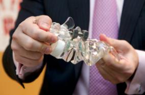
Summary: Dura is the fibrous membrane that covers the brain and spine. Perforations in the dura (durotomy) such as punctures, incisions or lacerations are often encountered during cranial and spinal surgery and require repair and sealing to prevent serious and occasionally fatal complications related to leakage of cerebrospinal fluid (CSF). Minimally invasive surgical (MIS) procedures of the brain and spine significantly limit direct repair of durotomies by traditional methods such as suture or staple closure, due to a combination of restricted working space and access vectors and limited field of view (constrained deployment), and the often-friable nature of the perforated dura. PatchClamp MedTech, Inc. is developing novel devices to enable rapid, safe, and immediate watertight repair and sealing of durotomies encountered during MIS and open craniospinal procedures. A low-profile, single-hand disposable applicator enables the surgeon to place a tissue or synthetic graft through a narrow MIS access port onto the inner surface of the perforated dura and secure it in place with a clasp released onto the outer dural surface. Prototypes using high resolution 3D printed bioabsorbable subcomponents and commercially available synthetic dural substitutes have provided proof-of-concept. In this proposal, we refine and test the efficacy of the device under a variety of frequently encountered MIS constraints in vitro and demonstrate a stable watertight seal in vivo. Ease of application, sealing efficacy and survey feedback from surgeons deploying the graft in this model will guide sequential modifications and scaling of the device. Aim 1. Assess mechanical attributes of device components and dural sealing performance in vitro in unconstrained access. Using clinically relevant burst pressure chamber models, we will assess the following attributes: easy loading and release of the graft from the applicator, atraumatic passage of the graft through the dural perforation with rapid re-expansion, precise release of the extradural clasp struts, elastic reformation of the struts against the membrane after release, measurement of the force applied by the struts to the external surface of the membrane, minimum strut force to maintain stability against lateral movement, and ability to unclasp and reposition or remove the graft after initial deployment. We will demonstrate that the deployed graft maintains a watertight seal under escalating pulsatile pressure in comparison to current dural closure techniques. Aim 2. Assess performance in vitro under constrained access conditions. We will assess visualization, access, deployment, and sealing efficacy compared to current dural closure techniques under constrained access models simulating MIS approaches using a lumbar soft tissue simulator with both a pressurized acrylic burst chamber as well as a pressurized spine laminotomy model. Surgeon input will guide design iterations. Aim 3. Demonstrate sealing performance in vivo. The refined prototype will be used to test device graft deployment and sealing compared to conventional dural closure in an acute pig laminectomy durotomy model. Successful completion will lead to preclinical IDE-enabling work in preparation for first-in-human trials.
Sponsor: Small Business Innovation Research (SBIR)


