Overview
Subarachnoid hemorrhage results from the bleeding of an artery around the base of the brain. It is the least common type of stroke, accounting for about 5 percent of all strokes.
Symptoms
For subarachnoid hemorrhage, the most common symptoms are the sudden onset of the “worst headache of my life” often with an associated stiff neck, and possible loss of consciousness.
If you or someone you know may be having a stroke, call 911 immediately.
Causes
The most frequent cause of subarachnoid hemorrhage is bleeding from an aneurysm. An aneurysm is a weakening and ballooning of a short portion of an artery (similar to a bubble on the side of an old hose).
Risk Factors
The factors that can increase a person’s risk of this type of stroke include high blood pressure, smoking and a family history of ruptured aneurysms.
Diagnosis
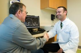
The doctors will need to make sure that the symptoms you are experiencing are due to a stroke. In order to do this, the doctors will find out more about you by asking you and your family about your medical problems and about the symptoms you are experiencing.
You will likely have an EKG to assess your heart rhythm and activity. The doctors will need to do a physical exam and draw some blood to send to the laboratory. Pictures of your brain will also be obtained in most circumstances. These images will help to determine if you are having an ischemic stroke (blockage without bleeding) or if you have bleeding in or around your brain (hemorrhagic stroke).
Pictures of your brain may be obtained using any of the following:
-
Computed tomography (CT scan): A CT scan may be normal if it is done soon after the onset of symptoms. A CT scan is the best test to look for bleeding in or around your brain. In some hospitals, a perfusion CT scan may be done to see where the blood is flowing and not flowing in your brain.
-
Magnetic resonance imaging (MRI scan): A special MRI technique (diffusion MRI) may show evidence of an ischemic stroke within minutes of symptom onset. In some hospitals, a perfusion MRI scan may be done to see where the blood is flowing and not flowing in your brain.
-
Angiogram: a test that looks at the blood vessels that feed the brain. An angiogram will show whether the blood vessel is blocked by a clot, the blood vessel is narrowed, or if there is an abnormality of a blood vessel known as an aneurysm.
-
Carotid duplex: A carotid duplex is an ultrasound study that assesses whether or not you have atherosclerosis (narrowing) of the carotid arteries. These arteries are the large blood vessels in your neck that feed your brain.
-
Transcranial Doppler (TCD): A TCD is an ultrasound study that assesses whether or not you have atherosclerosis (narrowing) of the blood vessels inside of your brain. A TCD can also be used to see if you have emboli (blood clots) in your blood vessels.
-
Echocardiogram: An echocardiogram is an ultrasound study of the heart. It can be done by placing the ultrasound probe on your chest (a transthoracic echocardiogram) or in your throat (a transesophageal echocardiogram). The purpose of an echocardiogram is to see if there are blood clots in your heart that may have led to your stroke.
Complications
Movement of the extremities is often impaired after a stroke. But the ability to move usually improves substantially during the first 30 days after the stroke and can continue to improve for up to 90 days. Recovery takes twice as long when there is serious impairment of movement. Most patients will have some remaining impairment of motion following their stroke.
Speech ability is also often impaired after a stroke. Speech improves in most patients. For those with mild speech impairment, improvement occurs within two weeks. For severe speech impairment it can take 10 weeks. Some patients continue to show improvement for months or years afterward.
Stroke can also interfere with your ability to think, remember, and pay attention to the world around you. Maximum improvement after the stroke usually occurs within three months but continued improvement may occur for a year.
Recovery
Movement of the extremities is often impaired after a stroke, but the ability to move usually improves substantially during the first months after the stroke and can continue to improve for some time thereafter. Recovery takes longer when there is serious impairment of movement. Most patients will have some remaining impairment of motion following their stroke.
Speech ability also is often impaired after a stroke. Speech improves in most patients. Some patients continue to show improvement for months or years afterward.
Strokes can be very different for different people, and the effects will vary depending on what part of the brain is injured. Because of this, it is not easy to generalize about the extent of improvement that you can expect. In addition, the patient’s outcome will depend on his/her general health. A healthy, active, young person will have a better outcome than an elderly, ill person.
If a person has only mild symptoms during the first few days after a stroke, they will generally be able to function better after recovery than a person who had severe symptoms after their stroke. Younger patients will have better recovery than elderly patients. The larger the area of damage to the brain, the less chance there is of getting full recovery.
When brain cells die as a result of a stroke, the body removes the dead cell debris. The brain function that has been impaired by the stroke can recover.
Undamaged brain areas near the area of the stroke may have impaired function as a result of the loss of normal communication with the damaged tissue. But after time, these regions resume their activity and some of the functions that were performed by the damaged area can be taken over by a neighboring healthy area of the brain and allow recovery to occur.
In the normal brain, an activity, such as pointing a finger, is the result of coordinated activity in many different parts of the brain. When a stroke damages one part of this network of activity, the remaining parts of the brain increase their activity in an effort to recover the function.
In addition, there are two brain hemispheres and when an area in one hemisphere is damaged, many patients start to use both sides of the brain to perform a task.
Today, good medical care allows the brain to heal after a stroke. Physical, occupational and speech therapy all play an important role in patients that have disabilities from their stroke. Family and other caregivers are vital to support the patient’s efforts toward recovery.
The brain has mechanisms to repair itself, but scientists are just beginning to understand them. The results from some experimental studies show that some medicines that stimulate brain cell activity or cause brain cells to grow can increase recovery from stroke. In other studies, introducing cells into the brain can promote recovery.
There are many reasons to be optimistic about meaningful recovery after a stroke.
Self Care
The risk of subarachnoid hemorrhage can be reduced by:
-
Treating high blood pressure (hypertension)
-
Stopping Smoking
-
Getting treatment for heart diseases
-
Maintaining a healthy lifestyle

