Overview
Gamma Knife does not actually involve cutting a tumor with a knife or scalpel. Instead the procedure, also known as stereotactic radiosurgery, uses high-energy gamma rays to pinpoint and destroy tumors or other brain abnormalities.
The UW Medicine Gamma Knife Center at Harborview offers the latest Gamma Knife technology, the Gamma Knife Perfexion and Perfexion Extend. This system uses 192 emitting sources of cobalt-60. Although the beam from each of these sources is relatively weak and does not damage tissue, when focused together, the combined beams can help destroy or contain the tumor while sparing nearby healthy tissue and other parts of the brain.
In most cases, it is a one-day, outpatient procedure that requires little or no anesthesia. A frame is fitted to the patient’s head. With the help of computerized imaging, our neurological surgery and radiation oncology team is able to map the exact location of the lesion, and then direct the beams at the tumor with extreme accuracy.
Gamma Knife is particularly effective in reaching small, localized tumors deep inside the brain that would otherwise be difficult to reach. Gamma knife radiosurgery is most commonly used for:
-
Brain tumors
-
Arteriovenous malformations (AVMs)
-
Trigeminal neuralgia "Tic Douloureaux"
-
Acoustic neuromas
-
Pituitary tumors
Procedural Details
Patients will usually be asked not to eat or drink anything for six hours before their Gamma Knife procedure. A Gamma Knife nurse will call before treatment to discuss specific instructions.
It is an outpatient procedure, usually completed in a single session, with the patient discharged home the same day. Patients should plan on spending the entire day at the Gamma Knife Center and arrange for a ride home.
Gamma Knife radiosurgery requires precise positioning. A specialized frame is fixed to the patient’s head to help aim the Gamma Knife at the right location in the brain and to keep the head steady and properly positioned during the procedure.
Image mapping and planning
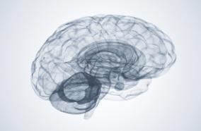
Imaging, which may include MRI, CT and/or a cerebral angiogram, is done after the frame is fitted and is used to locate the target area or areas. Once the targets are located, treatment planning software is used to create three-dimensional models of the brain. These models closely match the shape of the tumor or the affected area(s). Then a prescription dose is determined. This dose will be dependent upon the type of lesion or lesions, location, size and any prior therapy.
Treatment
During treatment, the patient is monitored by a video system and two-way intercom that allows the patient to talk with the physician during the procedure. When the procedure is completed, the head frame is removed and the patient is evaluated and prepared for discharge. Many patients return to their normal activities the following day.
It may take many weeks or even up to several years before the effects of Gamma Knife treatment become apparent. The patient’s progress is monitored through follow-up imaging studies.
Medications
Some patients experience persistent swelling after the treatment. Steroid medications may be prescribed to help treat this type of swelling.
Considerations
More than 500,000 patients have been treated worldwide with a variety of different types of tumors and AVMs. Patients with the following conditions may also be candidates for the Gamma Knife procedure:
-
Epilepsy
-
Chronic pain
-
Tremor
-
Patients who have had a recurrent brain tumor following surgery
A multidisciplinary team of neurosurgeons and oncologists will determine if Gamma Knife is the best option for each particular patient.
Treating children
Some brain lesions in pediatric patients are suitable for Gamma Knife. For our young patients, general anesthesia is used and patients go home the same day.
Consultation and referrals
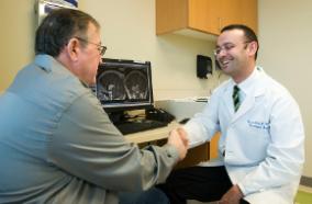
Typically, patients are seen in consultation and initially by a neurosurgeon or a radiation oncologist. In addition, our radiosurgery panel may review a case. After further evaluation—which can include MRI, functional MRI, CT, angiography or neuropsychological testing—the appropriate method of therapy is tailored for each individual's needs.
Effectiveness
Gamma Knife is used most effectively in the treatment of small intracranial tumors including:
-
Acoustic tumors
-
Meningiomas
-
Craniopharyngiomas and pituitary tumors
-
Arteriovenous malformations (AVMs)
Gamma Knife may be used in the treatment of lesions that cannot be reached by conventional surgery. It can be used along with other forms of radiation therapy, and with conventional surgery when complete removal of a tumor is not possible. It can be used in the treatment of recurrent tumors. Gamma Knife may also be used to help stop the growth of brain metastases from cancer.
The patient’s recovery depends on the size and type of abnormality, ranging from a few weeks to several years. Most abnormalities disappear gradually and others just stop growing. However, in some cases, the tumor or AVM might not respond to Gamma Knife therapy
Risks
Some side effects a patient may experience after Gamma Knife treatment include:
-
Nausea
-
Headaches
-
Skin irritation
-
Superficial lesions
-
Patchy hair loss
-
Neck stiffness
-
Dry mouth
-
Pain at the frame securing sites
Some patients experience a temporary swelling in the area near the treatment site. If the swelling persists, your doctor may recommend steroid medications.
Urgency
The decision to use Gamma Knife radiosurgery should be made after careful consideration with your doctor. The procedure may be used to treat brain tumors or other brain abnormalities, or it may be used to help control the spread of cancerous cells in the brain.

